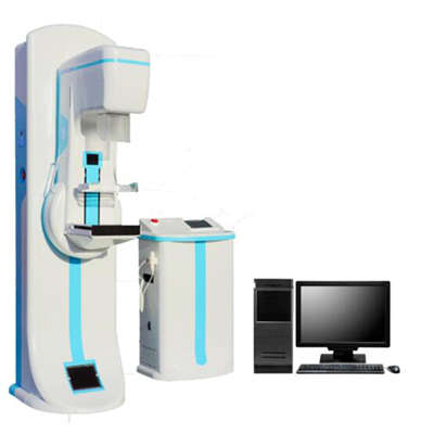
Digital Mammography System AM-500
I. Application
A mammogram is a special, low-dose X-ray technique used to take a picture of the breast, detecting and diagnosing any abnormal lumps or masses in breast tissue. It is one of the best tools for the early identification of breast cancer. With early identification, breast cancer can be cured while in the first stage, and recovery is more likely.
Description
II. Specification
| Item | Parameter | Remark |
| X-ray Generator | Generator Type: High Frequency Inverter 80kHz
Input Power: Single phase 220VAC, 50/60Hz Radiographic Ratings: Large Focal Point 20-35kV/10-510mAs Small Focal Point 20-35kV/10-100mAs Power Rating: 6.2kVA |
Self-developed and world advanced all-solid-state high frequency high voltage x-ray generator |
| X-ray Tube | Focal Spot Size: Dual Focus 0.1/ 0.3mm
Target Material: Molybdenum (Mo) Port Material: Beryllium (Be) High-speed anode drive: 2800 /10000rpm Target angle:10°/16° Anode Heat Storage: 210kJ (300kHU) Anode Cooling: Air cooling Filtration: Mo(0.03mm), Al(0.5mm) |
Model:IAE C339V
China tube for optional |
| Radiographic Stand | C-ARM: Vertical Movement: 590mm
Center of electricrotating C-arm Automaticreturn function by one key Rotations Degree: +90°~-90° Automatically released after the exposure pressure settingsdisplay Compression flexible, stepless speed. Max. pressure: 200N Max. travel: 150mm SID: 650mm |
Electric Isocentricrotating |
| Flat Panel Detector | Detector material: Amorphous silicon
Effective coverage of detector: 18x24cm Pixel matrix: 3072×1944 Limit of spatial resolution: 6.0Lp/mm DQE value: 70% dynamic range: 14bit digital output pixel size: 75μm High voltage Synchronizer trigger: BNC Output: Camera Link or Ethernet Working condition: 10℃-40℃ storage environment: -10℃-50℃ |
China Flat Panel Detector
24x30cm for optional |
| Bucky housing and movement device | Size: 374*304*65mm
Stepless speed regulating range: 0~6cm/s Movement range: 0.5~2cm Grid Size: 24x30cm Grid ratio: 5:1 Grid density: 30lp/cm Focal distance: 650mm |
|
| Image acquisition workstation | CPU≥Intel Core Duo 2.60GHz
Hardware≥250G high speed Hardware Memory≥2G Display card≥512MB high brightness high-contrast LCD,1280*1024 Pixel resolution Network interface Work-list DICOM3.0 transmission 100/1000 Gigabit Ethernet Software Imaging software packageDMOC V1.0 |
Configuration including Diagnose digital workstation
5M medical monitor for optional |
| Others | Line Voltage
220Vac±10%@25A,Single phase |
110V for optional |
III. Configuration
| No. | Item | Quantity |
| 1 | X-ray Tube | 1 |
| 2 | X-ray Generator | 1 |
| 3 | Gantry assembly | 1 |
| 4 | C-ARM | 1 |
| 5 | Bucky movement device | 1 |
| 6 | Flat panel detector | 1 |
| 7 | Image acquisition workstation | 1 |
| 8 | Review work station | 1 |
| 9 | Paddle switch | 2 |
| 10 | Exposal switch and connected line | 1 |
| 11 | Power wire | 1 |
| 12 | Grounded wire | 1 |
| 13 | Fuse | 2 |
| 14 | Operation manual | 1 |
| 15 | Maintenance Reference Manual | 1 |
- IV. Features
- Adopt specialized mammography flat panel detector digital imaging technology.
- Full size digital mammography x-ray imaging.
3. Unique adopt all-solid-state high frequency high voltage generator. This technology has got the PATENT IN THE USA.
Reviews
There are no reviews yet.